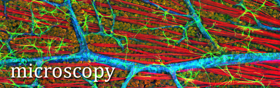
Mission Statement

The mission of NCMIR is to develop technologies to bridge understanding of biological systems between the gross anatomical and molecular scales and to make these technologies broadly available to biomedical researchers. NCMIR provides expertise, infrastructure, technological development, and an environment in which new information about the 3D ultrastructure of tissues, cells, and macromolecular complexes may be accurately and easily obtained and analyzed.
Read More >>
Read More >>
 Research Highlights
APEX2 Enhances Proximity Labeling and Electron Microscopy Imaging to Resolve Dispute about Regulation of the Inner Membrane Channel
A research team from MIT, Massachusetts General Hospital, and UCSD in late November 2014 published a study in Nature Methods describing an electron microscopy tag, APEX2, which they developed for live-cell proteomics and genetic probe-based labeling for light and electron microscopy. Use of this tag enabled them to resolve a conflict between competing mechanistic models of regulation of the inner membrane channel through which calcium is supplied to mitochondria. Read More >>
Automatic Segmentation of Organelles in Electron Microscopy Image Stacks: A New Workflow
Electron microscopy is a very useful technique to analyze the form, distribution, and functional status of key organelle systems in various pathological processes including those associated with neurodegenerative diseases. Significantly, it has been used to provide important new insights into the mechanisms underlying diseases such as Parkinson’s and Alzheimer’s diseases and glaucoma.Moreover, recent advances in EM instrumentation are fueling a renaissance in the study of quantitative neuroanatomy. Data obtained from techniques such as serial block-face scanning electron microscopy (SBEM) provide unprecedented volumetric snapshots of the in-situ biological organization of the mammalian brain across a multitude of scales. Read More >>
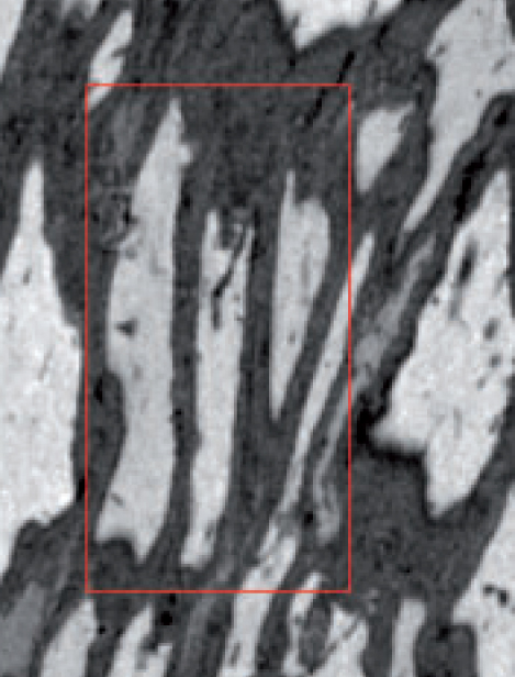
X-ray Microscopy Increases Accuracy and Efficiency of SBEM Imaging for Correlated Light/Electron Microscopy of Biological Specimens
The recently developed 3D method of serial block-face scanning electron microscopy is rapidly establishing itself as a powerful imaging approach in the biological sciences research community. This approach is proving of great value for 3D visualization of nervous system ultrastructure, particularly when details of synaptic and other subcellular elements must be located and quantified. It is also important for constructing connectomic models of regional brain circuits. In addition, SBEM is increasingly used to perform nanohistology on a wide variety of tissue systems, including the lung, liver, tendons, kidney, and cell cultures. Read More >>
Epik! A New Scientific Workflow for Electron Tomography
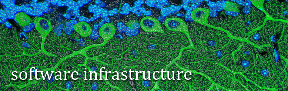
Computational researchers, regardless of their disciplines, need software tools that save time, optimize and scale up computations, produce results faster, create an extensible platform, foster collaborations, effectively communicate the underlying science, and enable others to replicate the results. Some of the most effective tools are based on scientific workflows. A workflow is a software application that solves a scientific problem. It’s composed of computational steps and data manipulation tools that can scale up to run on high-performance computers, distributed environments, and commercial cloud systems. Workflows are used in all stages of the data lifecycle: generation and acquisition, analysis, comparison, publication, and archiving. Read More >>
PRIME Directive: New Chemical Fluorophore Designed to Tag Cell Proteins for Imaging
Fluorescent proteins are used commonly to tag components of interest in living cells for subsequent imaging. But there’s a cost: Their dim fluorescence, rapid photobleaching, and large size limit their utility, and they can disrupt protein folding and trafficking and/or impair protein function.Chemical fluorophores, by comparison, tend to be considerably smaller, brighter, and more photostable, characteristics that allow them to perform better in advanced biological imaging modalities such as single-molecule tracking and super-resolution microscopies. However, it is much more challenging to target them to specific cellular proteins inside living cells because they cannot be genetically encoded and have to target proteins post-translationally inside a complex cellular environment. Read More >>
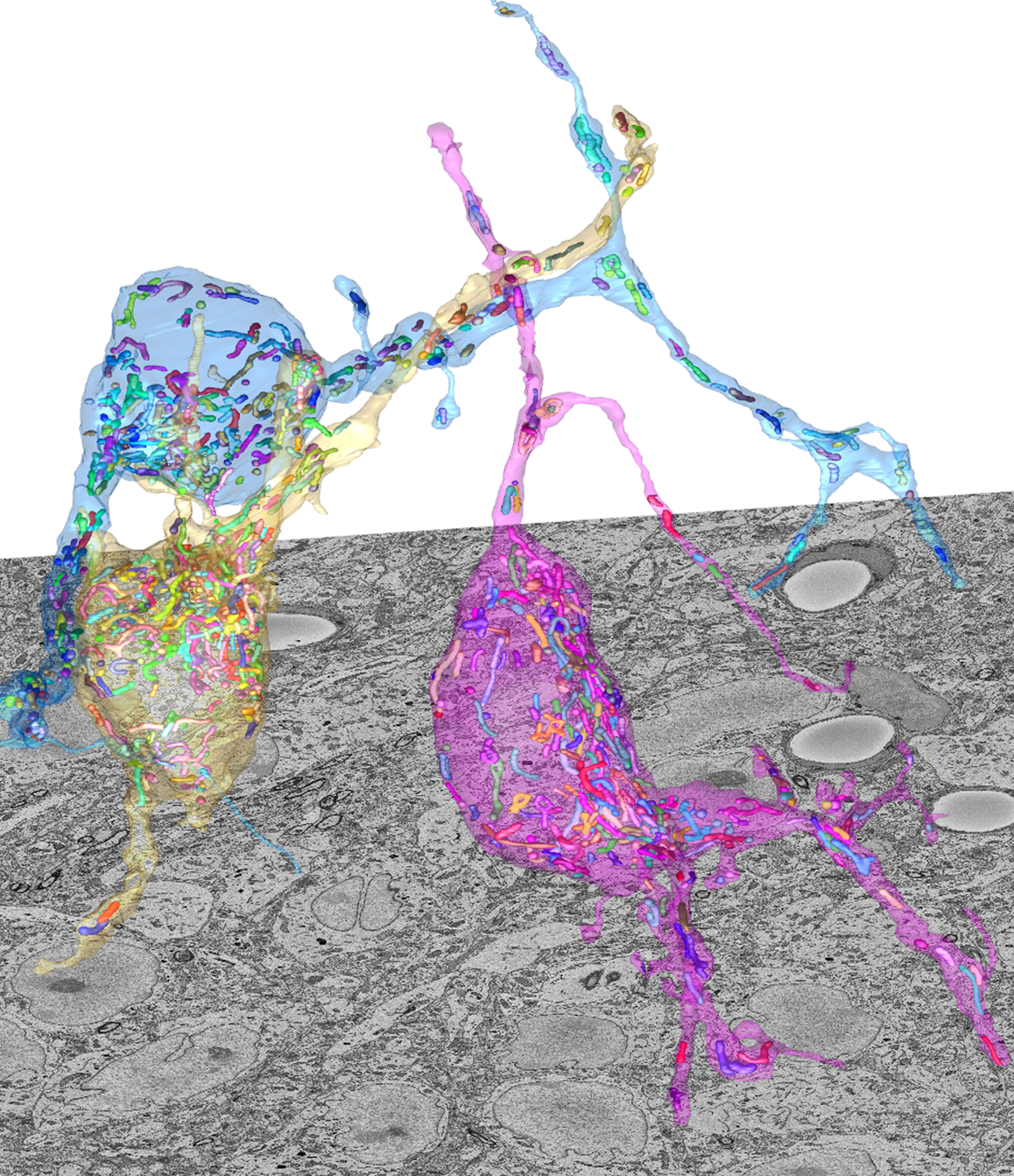
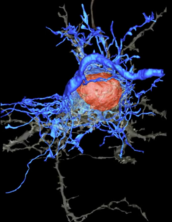
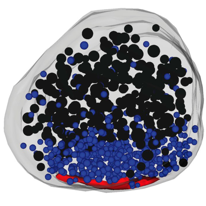
Astrocytes Devour Neuronal Mitochondria in a Newly Documented Process Called Transmitophagy
Scientific canon long held that when the tiny energy producers within cells, called mitochondria, become damaged or dysfunctional, these tiny organelles are degraded and recycled within the cell that produced them. But a paper appearing in the Proceedings of the National Academy of Sciences (PNAS) from The Johns Hopkins University and NCMIR scientists now shows that, instead, particularly in the long axons of the brain’s nerve cells, damaged mitochondria are transferred to nearby glial cells for elimination. This process, in effect, outsources a substantial portion of the neurons’ housekeeping. Read More >>
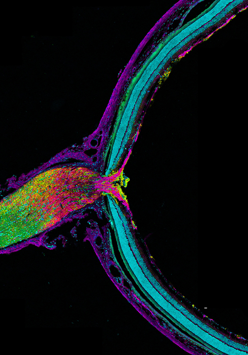
A New Model System for Neural Development Produces Nanoscale-resolution Animations that Inspires Music
Neural circuit development in mice is characterized by early exuberant innervation followed by competition and pruning to mature innervation topography. Several neural systems (e.g., the neuromuscular junction and climbing fiber innervation of Purkinje cells) have served as models to study neural development in part because they establish a recognizable endpoint of mono-innervation of their targets, and the presynaptic terminals are large and easily monitored. The calyx of Held (CH) innervation of its target, which forms a key element of auditory brainstem binaural circuitry, exhibits these same characteristic. Read More >>
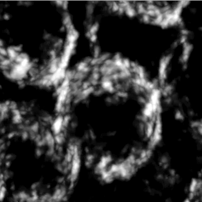
De-coating Caveolae: Understanding the Caveolar Protein Machinery in Endocytosis
Caveolae are flask-shaped invaginations with a diameter of 50-80nm in the plasma membrane of many types of mammalian cells. They are particularly abundant in fat cells, muscle cells, and cells that line blood vessels and serve as a source for clathrin-dependent endocytosis. Although they are likely to be important for cellular responses to mechanical stress, intracellular trafficking, and signaling events, the precise molecular mechanisms that determine how they form and carry out these functions have yet to be understood. Read More >>
Three Types of Microscopy Go To the Limit to Determine Ryanodine Receptor Calcium Release Channels in Cardiac Myocytes
A research team led by UCSD recently published results of their work using various kinds of microscopy to reveal the relative distribution of ryanodine receptors and caveolin-3 in mouse ventricular myocytes in the cytosol and near the cell surface. Ryandine Receptors are important to investigate because they comprise a class of intracellular calcium channels that mediate the release of calcium in various types of mammalian tissue like muscles and neurons. Read More >>
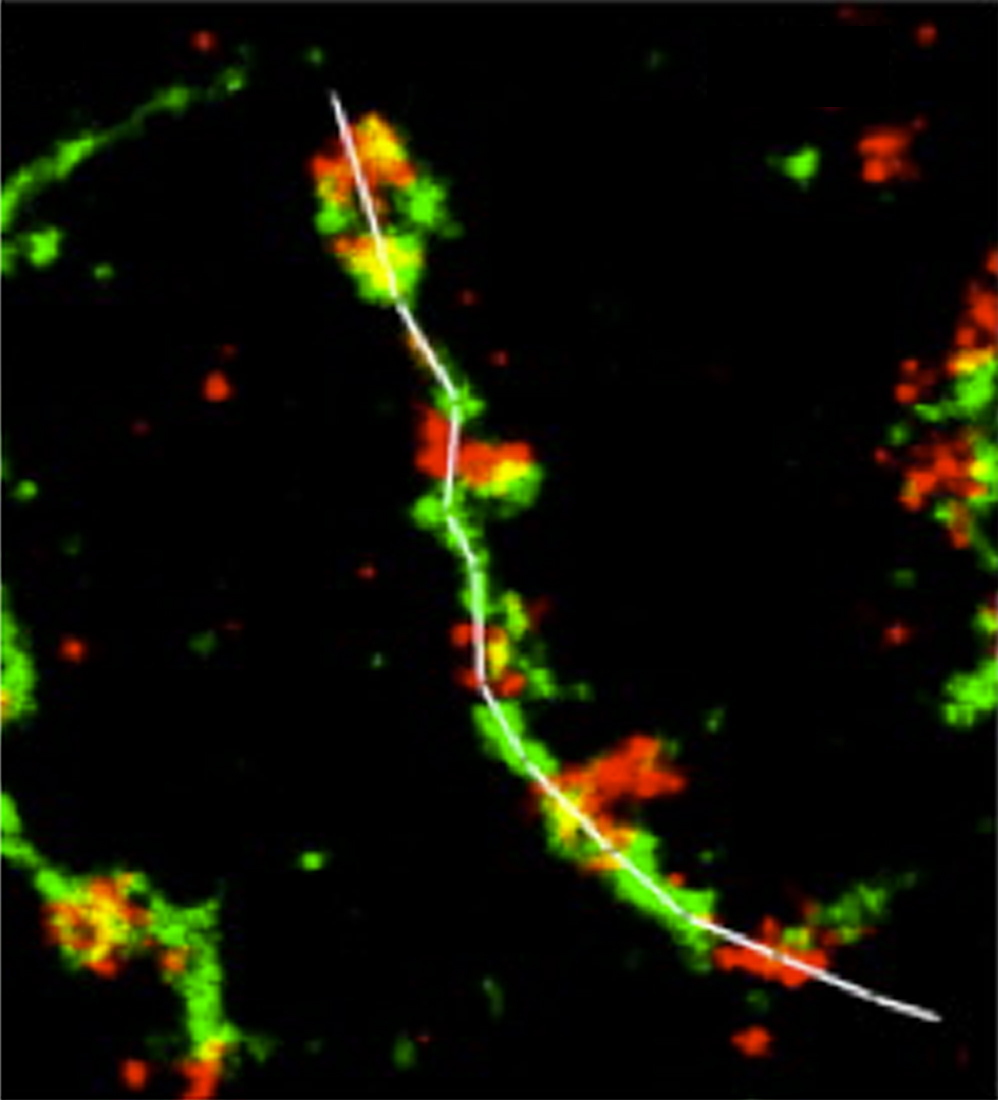
Liprin-α1/SYD-2 Determines Size of Electron-dense Projections in Presynaptic Active Zones in C. elegans
Fast synaptic neurotransmission relies on triggered release of neurotransmitters from synaptic vesicles after fusion with the plasma membrane. The release of synaptic vesicles is a highly regulated process of sequential events: First synaptic vesicles are recruited to the presynaptic active zone, then docked to the plasma membrane in a release-competent state, which guarantees their rapid release after the influx of calcium into the presynaptic terminal. Read More >>
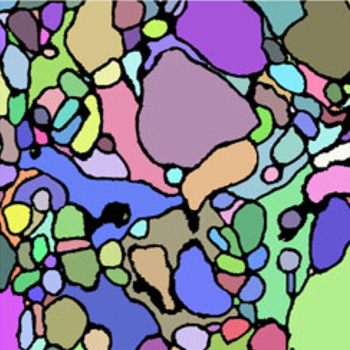
Big Data Analysis: Applying Electron Microscopy to Semi-automated Neuron Boundary Detection and Non-branching Process Segmentation
Reconstructing neural circuits is important to studying neural circuit connectivity and its behavioral implications. Understanding the differences between neuronal classes, patterns, and connections is critical to enable a more general understanding of how neural circuits process information. Electron microscopy is a useful method to determine the anatomy of individual neurons and their connectivity because it has a resolution that is high enough to identify features such as synaptic contacts and gap junctions, which define connectivity and, therefore, are required to reconstruct the neural circuit. But because the complexity and size – often approaching tens of terabytes -- of this data make for very difficult and labor-intensive interpretation, new segmentation techniques for identifying neurons in these data sets are needed. Read More >>
Efficient, Scalable, and Cost-effective 3D Microscopy Image Segmentation Using a Microlabor Workforce
In an applications note published in Bioinformatics, a team of researchers from the National Center for Microscopy and Imaging Research at UCSD described a new scalable, semi-automated approach to segment 3D structures revealed in serial block-face scanning electron microscopy (SBEM) images. The team used the Dual Point Decision Process (DP2) to divide the segmentation problem into discrete tasks distributed to a large pool of individual workers. Read More >>
Adenovirus Protein may be a Key to Strategic New Cancer Therapies
A team of scientists led by Clodagh O’Shea’s group at The Salk In100stitute for Biological Studies in collaboration with NCMIR scientists (Mark Ellisman, Tom Deerinck, Andrew Noske) and Dr. Roger Tsien and his laboratory suggest that cold viruses could serve as a valuable tool in the fight against cancer. Adenovirus, a type of cold virus, has developed proteins that allow it to hijack a cell's molecular machinery particularly those involved in growth, replication and cancer suppression. Read More >>
 In The News
NCMIR’s own Picture Wizard Tom Deerinck featured in the January issue of “The Scientist” magazine
NCMIR researcher Tom Deerinck was profiled by writer Jef Akst in the January 2015 issue of the magazine “The Scientist”. Deerinck is a completely unique individual with a novel career history of creating high works of arts from his impeccable scientific studies. The magazine profile details how he came to work when he was 20 years old under the tutelage of distinguished professor Mark Ellisman at UCSD in 1978 as well as how his passion for the scientific process and imaging on state-of-the-art microscopes endures. Read More >>
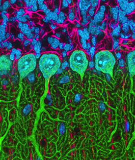
NCMIR researcher captures second place in the 11th annual Olympus BioScapes International Digital Imaging Competition
Winners of the annual Olympus BioScapes Competition were announced December 16, 2014, and NCMIR researcher Thomas Deerinck garnered second place for his triple labeled confocal fluorescence image of cerebellar Purkinje neurons in rodent brain. The competition, which is widely recognized as one of the preeminent photography competitions focusing on microscopic imaging in the life sciences, attracted over 2500 entries from 70 different countries throughout the world. Read More >>
UC San Diego and NCMIR Researcher Roger Tsien is awarded the first annual Golden Goose Award from Congress
Roger Tsien, PhD, Professor of Pharmacology at UC San Diego and Nobel laureate for Chemistry 2008, for work related to NCMIR at UC San Diego, received the Golden Goose Award in September 2012. This award “showcases researchers who pursue oddball topics that eventually lead to significant health and economic benefits, the awards were created by a coalition of science organizations (including AAAS, publisher of Science Insider) Read More >>

The Microscopy Society of America awards the Maser Award to NCMIR’s own Gina Sosinsky
At the 2012 Microscopy & Microanalysis meeting held in Phoenix this July, Gina Sosinsky, Assistant Director of NCMIR and a UCSD Professor-In-Residence in Neurosciences was awarded the Morton D. Maser Ward for Distinguished Service to the Microscopy Society of America. This major society award recognizes outstanding volunteer service to the Society. Read More >>

NCMIR at the MET
NCMIR Commissioned to Provide Artwork for the New Medical Education and Telemedicine Center at UCSD
La Jolla, CA , December 2011- Researchers from the National Center for Microscopy and Imaging Research (NCMIR) have been charged with providing science-based artwork for the public areas of the new +100,000 square foot Medical Education and Telemedicine Center on the campus of the School of Medicine at UCSD. The new center will be a hub of learning that incorporates state-of-the-art design and technology to prepare medical students to become physicians and innovators of tomorrow. It will also be used for physicians to learn new skills utilizing the latest advances in medical and surgical technology, such as surgical robotics. Read More >>