Research Highlight
NCMIR Researchers Help Create a Powerful New Probe for Light and Electron Microscopy
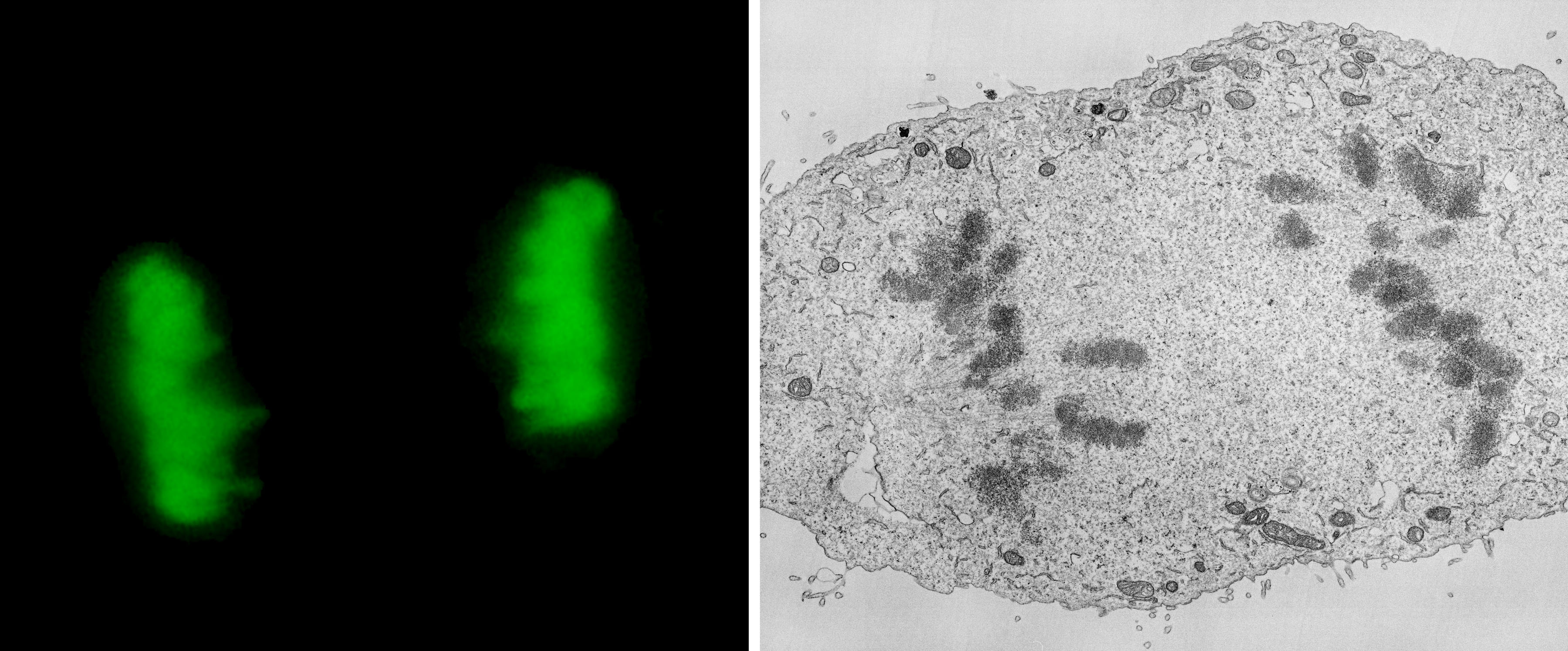
La Jolla, April, 2011 —In the April 5th issue of PLoS Biology, NCMIR scientists, in a collaboration with a team led by Nobel laureate Roger Tsien, introduced a new fluorescent probe for correlated light and electron microscopy that overcomes many of the limitations previously encountered by researchers seeking to image cells and tissues at high resolution. This probe, termed “miniSOG” (for mini singlet oxygen generator) was genetically engineered from a portion of the native protein phototropin-2 from the plant Arabidopis thaliana and can be used much like the genetically encodable family of fluorescent proteins obtained from jellyfish that have revolutionized modern light microscopy. However, this new probe has the added capability to act as a high-resolution protein label for electron microscopy as well. In a process referred to as fluorescence photo-conversion, singlet oxygen produced by the light-stimulated excitation of miniSOG is used to create a reaction product in a highly efficient manner that is visible by electron microscopy. The advantage of electron microscopy is that it has much higher spatial resolution than light microscopy. The result is that extraordinarily detailed, three-dimensional images of a wide range of biological specimens with highly specific labeling can now be obtained. Both Tsien and Ellisman agree that the development of miniSOG may prove as useful and revolutionary for electron microscopy as GFP has been for light microscopy, and miniSOG is already being used in a whole host of collaborative research projects. The NCMIR team was headed by Thomas Deerinck and Mark Ellisman and included members of the Tsien lab as well as those of Professor Yishi Jin at UCSD and Michael Davidson of Florida State University.
Citation: Shu X, Lev-Ram V, Deerinck TJ, Qi Y, Ramko EB, et al. (2011) A Genetically Encoded Tag for Correlated Light and Electron Microscopy of Intact Cells, Tissues, and Organisms. PLoS Biol 9(4): e1001041. doi:10.1371/journal.pbio.1001041
Press Release April 5, 2011 - Rejuvenating Electron Microscopy