Research Highlight
A New Model System for Neural Development Produces Nanoscale-resolution Animations that Inspires Music
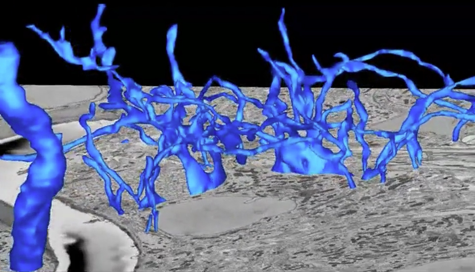
Figure Caption: Segmentation of the calyx of Held in a serial block-face scannig electron microscopy volume from a developing mouse brain.
January, 2014 La Jolla -- Neural circuit development in mice is characterized by early exuberant innervation followed by competition and pruning to mature innervation topography. Several neural systems (e.g., the neuromuscular junction and climbing fiber innervation of Purkinje cells) have served as models to study neural development in part because they establish a recognizable endpoint of mono-innervation of their targets, and the presynaptic terminals are large and easily monitored. Calyx of Held (CH) innervation of its target, which forms a key element of auditory brainstem binaural circuitry, exhibits these same characteristics. To investigate CH development, this team applied serial block-face scanning electron microscopy (SBEM) for the first time to neural development and thereby accomplished the first time series for 3D ultrastructural analysis of neural circuit formation. SBEM is a new technology that enables visualizing the wiring of the brain in 3D with a resolution of several nanometers -- about the size of a protein molecule. This approach revealed a growth spurt of added apposed surface area (ASA) in mice at postnatal day 3 and an initial rapid phase of growth and competition that resolved to monoinnervation in two-thirds of cells within three days. This growth occurred in parallel with an increase in action potential threshold, which the team projects may mediate selection of the strongest input as the winning competitor. ASAs of competing inputs were segregated on the cell body surface. These data suggest mechanisms to select “winning” inputs by regional reinforcement of postsynaptic membrane to mediate size and strength of competing synaptic inputs.
The team published their results in three consecutive issues of The Journal of Neuroscience: August 7, 14, and 21, 2013. The covers of those issues also featured the team’s work. In another first – for the journal – these cover image were taken from video animations rather than still images. The videos were created by Dr. George Spirou and colleagues at the West Virginia University (WVU) Center for Neuroscience. The first cover depicts the CH partially extracted from the image volume. The CH, located in the auditory central nervous system, is the largest nerve ending in the mammalian brain. This cover shows a nanoscale-resolution image from SBEM of the developing auditory brainstem during the first few days of postnatal mouse development. Such images are huge. One image volume can require up to 2 terabytes of computer storage space. New data sets that are at least triple this size are being collected by Spirou’s group at WVU using microscopes at the National Center for Microscopy and Imaging Research led by Mark H. Ellisman at UCSD.
The era of really big data has come to biology through studies of brain structure. It has been made possible by advances in computing speed, but the demand for even faster computers and more rapid-access storage of files of this size is relentless. These data will help us understand more about what’s happening within the brain as it develops. In particular, SBEM and related new technologies are enabling new ways to look at brain-based disorders, such as epilepsy, schizophrenia, and depression. Eventually, as image volumes continue to grow in size, we will be able to determine where the human brain’s wiring went wrong and understand the neural basis for these pathologies. In another likely first for scientific videos, each includes an original score by musician Bill Mallers. When Mallers saw the first video, he was really impressed by the visuals and asked if he could compose some music. Each score has a very different sound that reflects his interpretation of that video. As he explained it, the purpose of the music is to draw the viewer through the visual scenes.
(This article drew from a news release written and published 8/8/2013 by the West Virginia University Robert C. Byrd Health Sciences Center.)
Citation: Paul S. Holcomb, Brian K. Hoffpauir, Mitchell C. Hoyson, Dakota R. Jackson, Thomas J. Deerinck, Glenn S. Marrs, Marlin Dehoff, Jonathan Wu, Mark H. Ellisman, and George A. Spirou, Synaptic Inputs Compete During Rapid Formation of the calyx of Held: A New Model System for Neural Development, The Journal of Neuroscience, 2013, 33:12954- 12969; doi: 10.1523/JNEUROSCI.1087-13.2013.
A smartphone QR code is printed next to the study in the journal and links to the video when scanned.