Research Highlight
New insights into mitochondrial structure during cell death.
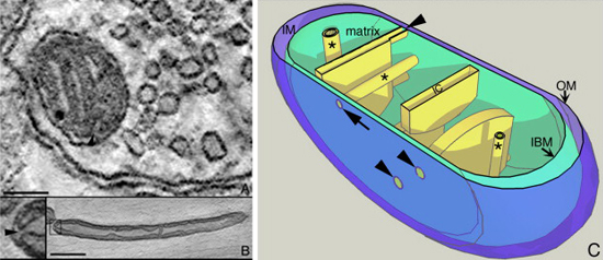
December 2009 — In this research effort, NCMIR neuroscientists and investigators at the Burnett School of Biomedical Sciences, College of Medicine, University of Central Florida, examined the pivotal role mitochondria play in the cascade of events associated with cell death pathways involved with several forms of neurodegeneration. NCMIR’s ability to successfully slice through an electron tomographic volume to image fragmented mitochondria revealed cristae vesiculation, cristae degradation and swollen endoplasmic reticulum.
This study confirmed that in the Bax/Bak-dependent pathway of apoptosis, the release of cytochrome c from mitochondria is a consequence of two carefully coordinated events: opening of crista junctions triggered by OPA1 oligomer disassembly and formation of outer membrane pores. Both steps are necessary for the complete release of pro-apoptotic proteins. The remodeling of mitochondrial structure accompanies this pathway, including mitochondrial fission, and cristae and crista junction alterations. There is remaining controversy surrounding the timing of certain remodeling events and whether they are necessary early events required for the release of pro-apoptotic factors or are simply a downstream after-effect.
In this paper NCMIR scientists thoroughly analyze the current knowledge of mitochondrial remodeling during cell death and discuss what structural alterations occur to this organelle during neurodegeneration, focusing on the higher resolution structural correlates obtained by electron microscopy and electron tomography.
Related Publications
G. Perkins, E. Bossy-Wetzel, M.H. Ellisman, New insights into mitochondrial structure during cell death, Exp Neurol 218 (2009) 183-192.