Research Highlight
EM Tomography Emerges as a Useful Tool for Studying the Nervous System
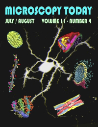
July 2003 — These images, featured on the July 2003 cover of Microscopy Today, demonstrate the emerging importance of electron tomography as a research tool, particularly for studying the nervous system.
The center image is a projection through an optical series recorded on a 2-photon microscope of a medium spiny neuron from the nucleus accumbens of the mouse filled with Lucifer Yellow.
The images surrounding the spiny neuron are reconstructions using single axis electron tomography and then surface rendered. Starting in the upper left-hand corner and proceeding clockwise is mitochondria (image by Guy Perkins), a synaptic complex from the rat hippocampus, a synaptic complex from the frog vestibular hair cell, a node of Ranvier from a mouse dorsal root peripheral nerve, a branched spiny dendrite from a rat cerebellum, and an unbranched spiny dendrite from rat neostriatum. The sizes of these structures range from 40 nm to 20 µm.